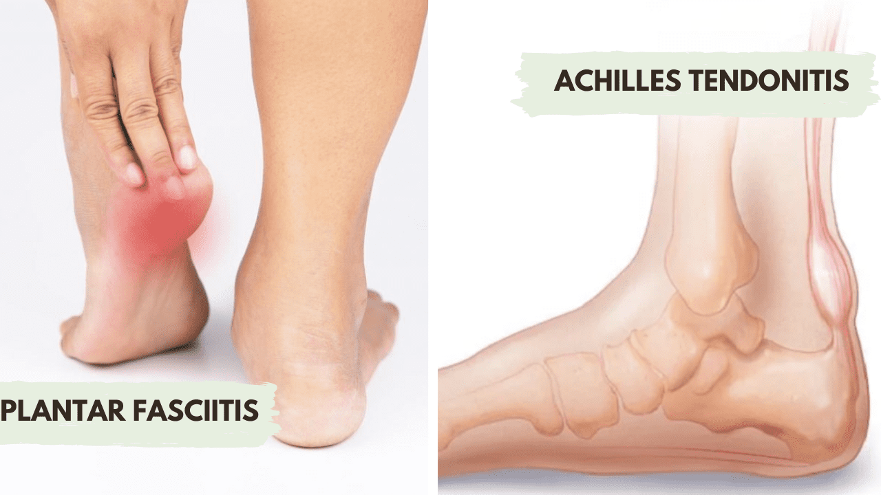
I came across a rather infrequent but recurrent situation that appears to confuse some of those managing workers’ compensation claim files. This involves pain in the distal lower extremity and the confusion is the difference between plantar fasciitis and Achilles tendonitis.
Both of these diagnoses are noted to be causative of distal lower extremity and or heel pain. However, both of these specific pathologies are very specific and different disease processes with their own unique characteristics. One needs to point out these differences so that the most appropriate diagnosis can be made, and a determination relative to the noted pathology being a function of the reported identified injury.
Plantar fasciitis is a painful situation noted on the bottom of the heel and can extend into the arch of the foot. This area of the foot is the plantar aspect, and the structure involved is the fascia attached to the calcaneus and the base of the toes. This structure supports the contents of the foot, particularly the tendons.
The symptoms include sharp pain, pain identified with the first steps after getting out of bed in the morning, and with direct palpation using the examiner’s thumb pushing at the area of the anterior aspect of the calcaneus. In more advanced cases, this type of palpation is particularly painful. Generally, this inflammatory process (“itis”) Is a chronic finding oftentimes lasting more than a year.
The causes of plantar fasciitis include overuse or a repetitive strain type scenario, poor foot mechanics or biomechanics, ordinary disease of life degenerative changes, obesity, and running or jumping on hard surfaces. Each of these activities can result in an inflammatory process of the plantar fascia.
Achilles tendonitis is pain noted on the posterior aspect of the heel, where the Achilles tendon attaches to the calcaneus. The Achilles tendon (noted to be the largest tendon in the body) connects to the gastrocnemius muscle to the heel enabling plantar flexion which occurs during walking or running.
The causes of Achilles tendonitis include overuse or repetitive strain, increase in exercise intensity or frequency, pork calf muscle flexibility or strength, age-related degenerative changes, or running or jumping on a hard surface.
As you can see, the causes of both of these diagnoses are essentially the same. Each of the structures noted is attached to the calcaneus. This is a primary reason for the noted confusion of these specific diagnoses. The pathologic response to the identified insult would include, from an anatomic perspective, irritation, inflammation, and a painful situation.
Thus, the question then becomes how do you discern the difference between these two diagnoses? The answer is location. Plantar fasciitis will be tender on the bottom of the foot and Achilles tendonitis will be painful in the posterior aspect of the foot.
The next issue is, are either of these diagnoses a function of a specific isolated event? Given that each of these diagnoses are by definition chronic in nature, represent long term changes, and are a function of a repetitive micro insult, there needs to be specific objectification as to why this would be a function of a specific event to make this a compensable injury. Otherwise, from an evidence-based medicine perspective, these are chronic, ordinary disease of life findings are not a result of a specific compensable injury.
When reviewing the clinical records, the progress note must include a detailed/comprehensive clinical evaluation, one that includes a particularly specific clinical history, a markedly detailed physical examination of the lower extremity, and proper diagnostic imaging studies.
Depending on the severity, plain radiographs of the foot should be obtained. Osseous changes including traction spurs, a thickening of the plantar fascia, and other degenerative changes can be seen. If there is still a question, an MRI of the distal lower extremity is utilized to decide the presence of such pathology. It should be noted that MRI will be used if non-surgical interventions have not ameliorated the symptomology.
The treatment ranges from rest, ice, elevation, and non-steroidal medications through injection protocols and potential surgical intervention. As you can see, establishing the correct diagnosis, ascertaining the correlation of this pathology to the compensable event, and ensuring a comprehensive clinical assessment are provided is essential in managing a case where either of these diagnoses are alleged to have been a function of a specific compensable event.

